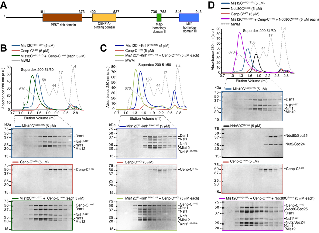
Figure 1. Cenp-C1–400 binds Mis12C.
(A) Schematic depiction of the domain organization of human Cenp-C. (B) Size-exclusion chromatography elution profiles and SDS-PAGE analysis of recombinant Mis12CNsl1(1–227) (upper panel), recombinant Cenp-C1–400 (middle panel), and their stoichiometric combination (lower panel). Complex formation is indicated by a shift in the elution profile of Cenp-C1–400 and Mis12CNsl1(1–227) and their appearance, in stoichiometric amounts, in early elution volumes. (C) As in B, but with Mis12CFL-Knl12106–2316 instead of Mis12CNsl1(1–227). The middle panel is the same as in B. (D) Incorporation of the Ndc80Bonsai complex in the Cenp-C1–400-Mis12CNsl1(1–227) complex. The upper panel and lower middle panel are the same as in B.

