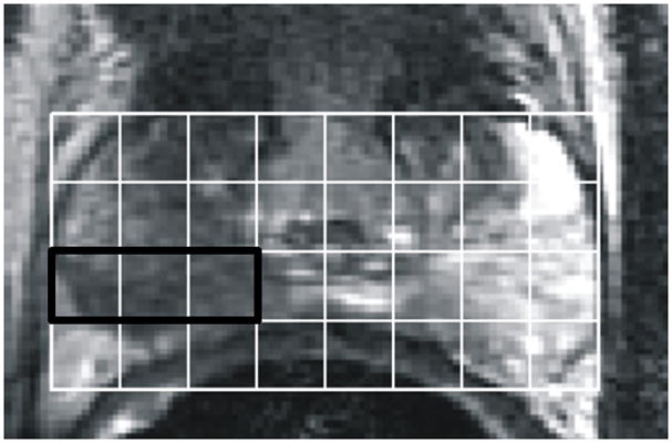
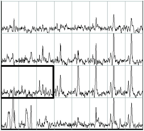
Figure 1.


Example of pre-operative proton MRI/MRSI data in a patient with prostate cancer (biopsy Gleason score = 3+4, PSA= 5.4, clinical stage = T2a). The figure contains single slice data extracted from a three-dimensional data set. A) T2-weighted image with grid indicating division of tissue into volume elements (voxels). B) Corresponding MRSI spectra for each voxel in the grid. The bold box includes voxels comprising the MRSI index lesion which have reduced citrate and elevated choline. On the left side of the peripheral zone (right side of image), high levels of citrate indicate healthy glandular tissue. Tumor was confirmed by surgical pathology. 3D-MRSI required 17 minutes and yielded spatial resolution of 6.25 mm in all three dimensions with an interpolated voxel size of 0.12 cm3. Voxels were classified as suspicious for cancer if [Cho+Cr]/Cit (CC/C) was at least 2 standard deviations above the average healthy ratio for the peripheral zone (PZ) 30.
