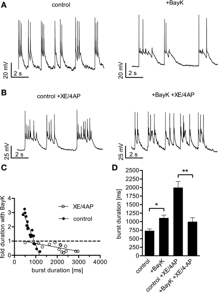
Fig. 6.
Effect of LTCC activation on neuronal activity depends on the extent of depolarization. When BayK was added to neurons displaying a burst firing mode, the duration of the depolarization wave was increased or decreased, depending on the length of the burst before DHP application. A: recordings from a burst firing neuron under control conditions and in the presence of BayK reveal a prolonging effect of LTCC activation on the depolarization waves. B: recordings from a bursting neuron treated with the potassium channel blockers XE 991 (XE, 10 μM) and 4-aminopyridine (4AP, 100 μM) showing particularly long-lasting depolarization waves that were shortened after addition of BayK. C: plot of fold change induced by BayK against burst duration in otherwise untreated cells (solid circles) and in cells after coapplication of XE and 4AP (open circles). The solid lines were generated by linear regression analysis yielding r2 = 0.89 for data obtained under control conditions and r2 = 0.50 for data obtained in the presence of XE/4AP. The intersection of the regression line for control data with the dashed line representing unaltered burst duration is at 924 ms. D: summary of the overall enhancing effect of BayK on burst durations in otherwise untreated neurons, the prolonging effect of XE/4AP, and the shortening effect of BayK on burst discharge in the presence of these potassium channel blockers. Bars represent the means ± SE of the median burst duration. The n value was 17 cells for burst activity under control conditions and 14 cells for burst activity in the presence of XE/4AP. * and **Statistically significant difference of P < 0.05, and P < 0.01, respectively, as determined by a Mann-Whitney test.

