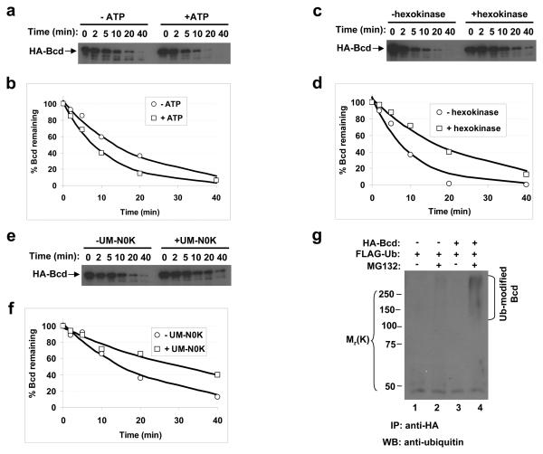
Figure 2. Bcd is ubiquitinated.
(a, c and e) Time course of Bcd degradation in embryonic extracts with or without ATP addition (a), ATP depletion (c) or UM-N0K addition (e). Hexokinase (together with glucose) was used to deplete ATP. For each experiment, aliquots were taken from the reaction tubes at the indicated time and HA-Bcd was detected by Western blotting.
(b, d and f) Fraction of remaining Bcd protein (from panels a, c and e) plotted against reaction time. In each case the lines represent the exponential fitting to the experimental data. Based on three independent experiments, the estimated half-life of Bcd is increased by 1.93 ± 0.35 fold in presence of UM-N0K.
(g) Bcd ubiquitination detected in cells. Whole cell extracts from HEK 293T cells were immuno-precipitated (IP) by an anti-HA antibody and analyzed by Western blotting using the anti-ubiquitin antibody. Ubiquitin-modified Bcd products are marked, and molecular weight standards are shown on the left.

