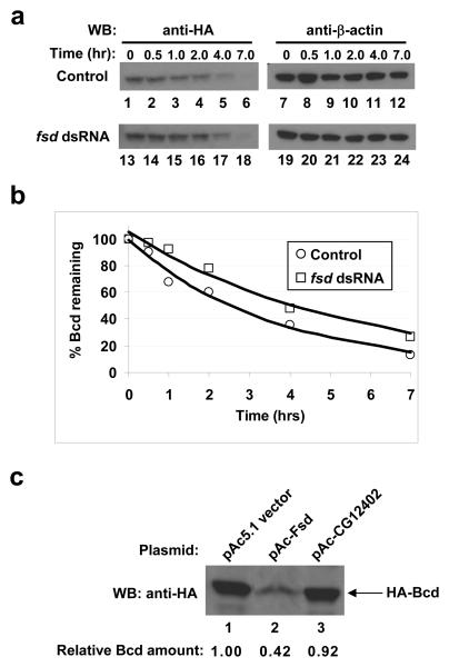
Figure 3. Fsd plays a role in Bcd protein degradation.
(a) CHX assay in HA-Bcd-expressing stable cells. Cells were treated first with fsd dsRNA, then with CHX, and harvested at indicated time (of CHX treatment) for Western blotting to detect the total amount of Bcd in cells (left panels). β-actin (right panels) represents loading control.
(b) Fraction of remaining Bcd protein (from panel a and normalized by β-actin intensities) plotted against time after CHX addition. The lines represent exponential fitting to the experimental data.
(c) Over-expression of Fsd enhances Bcd degradation in Drosophila S2 cells transiently transfected with the HA-Bcd expressing plasmid. For each lane, the loading amount had been adjusted by the β-galactosidase activities (measuring transfection efficiency) expressed from a lacZ control plasmid.

