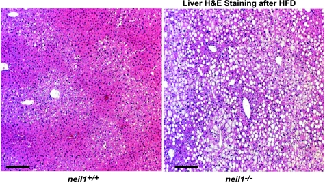
Fig. 6.
Hepatic lipid accumulation. Liver sections from HFD-fed neil1+/+ and neil1−/− mice were stained with hematoxylin and eosin (H & E). Neil1−/− animals displayed dramatic accumulation of hepatic lipids, seen as white droplets, following a 5-wk HFD feeding. A blinded assessment of liver damage revealed neil1−/− mice to have moderate to severe hepatic steatosis, whereas neil1+/+ mice displayed only mild steatosis. Images are representative of 5–6 animals/group, and the bar represents 25 μm in both images.

