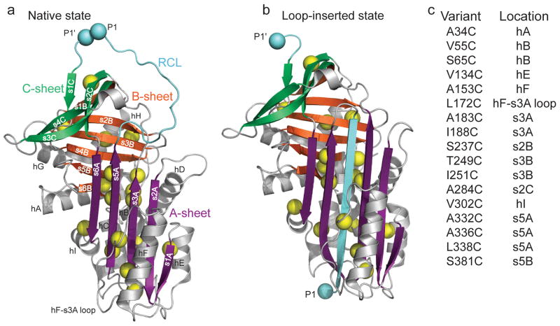
Figure 1.
Design of cysteine mutations to probe low denaturant-induced strand opening in the serpin α1AT. Comparison of structures of (a) native (PDB 1QLP)31 and (b) cleaved ‘loop-inserted’ (PDB 1EZX)2 states of α1AT. The two light blue spheres, labeled P1 and P1′, correspond to the protease cleavage site in the RCL. Sites mutated to cysteine are indicated by yellow spheres centered on the Cβ. (c) Positions of cysteine substitutions and their secondary structural context. Structures in this and other figures were prepared using PyMol (http://www.pymol.org).

