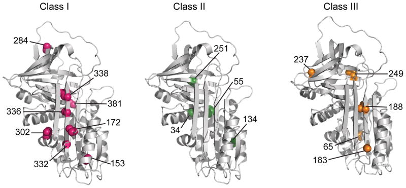
Figure 3.
Structural context of the cysteines belonging to the three classes with varying extents of PEGylation. Class I (pink), II (green), and III (orange) exhibit 100%, ~50% and <20% PEGylation, respectively, in the low GdmCl intermediate state of α1AT. The seventeen residues investigated in the current study are shown on the native structure of α1AT (PDB ID 1QLP)31.

