
Figure 2.
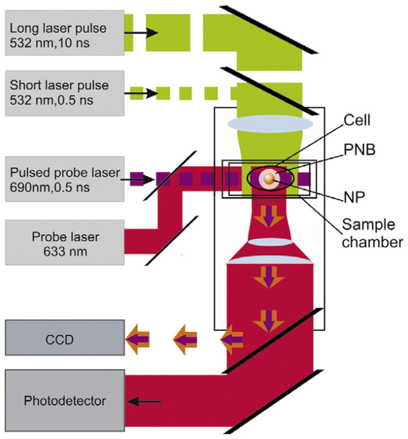
Experimental setup: single gold NPs in water or individual cells in the sample chamber were mounted on the stage of an inverted optical microscope; PNB generation was provided by focused single pulses (532 nm, 0.5 ns); a pulsed probe laser (690 nm, 0.5 ns) provided time-resolved optical scattering imaging of the PNB and a continuous probe laser (633 nm, 1 mW) provided monitoring of the optical scattering of PNBs though their time responses. An additional pulsed laser (532 nm, 10 ns, 1 mJ cm−2) was used for excitation of fluorescence in the cells.