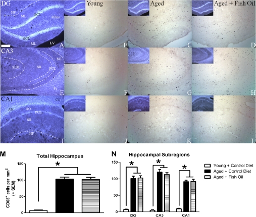
Figure 8.
Density of activated microglia is increased in hippocampus of aged rats. 4′,6-Diamidino-2-phenylindole dihydrochloride (DAPI) staining illustrating anatomical landmarks in the dentate gyrus (A), CA3 (E), and CA1 (I) of young rats. Representative photomicrographs obtained from young (B, F, J) and aged rats (C, G, K), and aged rats fed a fish oil–enriched diet (D, H, L) demonstrating CD68-immunoreactive cells (putative microglia) in the dentate gyrus (B–D), CA3 (F–H), and CA1 (J–L) of the hippocampus. Inset (B–D, F–G, and J–L): DAPI counterstain corresponding to each image shown. CD68+ cells were rarely observed in young rats, but density was significantly increased in aged rats regardless of diet (M and N; *p < .001 vs young control). Among aged rats, the density of CD68+ cells was significantly greater in the CA3 subregion relative to the CA1 (p < .001). Scale bar at bottom left in A is 250 μm. All images were acquired at the same magnification. Alv = alveus; CC = corpus callosum; Fi = fimbria; GCL = granule cell layer; HF = hippocampal fissure; LV = lateral ventricle; ML = molecular layer; PCL = pyramidal cell layer; SLM = stratum lacunosum-moleculare; SO = stratum oriens; SR = stratum radiatum.

