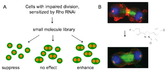
Figure 2. A pathway screen results in small molecules that target the Rho pathway in cytokinesis.
A. A small molecule/RNAi modifier screen. Cells with two nuclei are a consequence of failed cytokinesis and the readout in the screen. (Whole cells are cartooned in green, DNA in orange). B. Small molecules from the screen inhibit the accumulation of phospho-myosin at the cleavage furrow, a key function of the Rho pathway. Immunofluorescence images show Drosophila Kc167 cells where phospho-myosin (red), tubulin (green) and DNA (blue) have been visualized. Note the decrease of phospho-myosin at the furrow in compound-treated cells. Adapted from (7).

