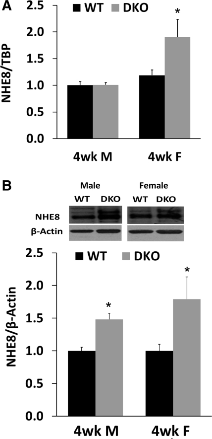
Fig. 4.
NHE8 expression in 4-wk-old NHE2X3 DKO mice. A: NHE8 mRNA expression in 4-wk-old male and female mice. RNA was isolated from the intestinal mucosa of 4-wk-old male and female wild-type mice and NHE2X3 DKO mice. Real-time PCR was performed using mouse-specific NHE8 and TBP primers. The changes in NHE8 gene expression are analyzed by the comparative cycle threshold (Ct) method. Data are means ± SE for each age group. *P ≤ 0.01 for NHE2X3 DKO mice vs. wild-type mice. Thirty wild-type mice (15 males, 15 females) and 29 NHE2X3 DKO mice (14 males, 15 females) were used. B: NHE8 protein expression in 4-wk-old male and female mice. BBMV was isolated from the intestinal mucosa of 4-wk-old male and female wild-type mice and NHE2X3 DKO mice. Western blot was used to detect NHE8 and β-actin protein abundances in BBMV preparations. The expression of NHE8 protein is calculated by the optical density of the NHE8 band over that of the β-actin band. Data are means ± SE for each gender group. *P ≤ 0.01 for NHE2X3 DKO mice vs. wild-type mice. Three independent experiments were performed. Inset: corresponding Western blot images.

