Fig. 4.
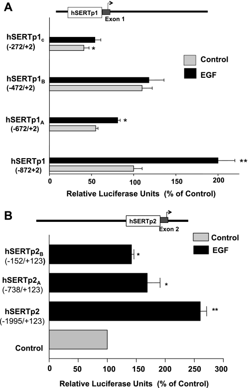
EGF-responsive regions of hSERTp1 and hSERTp2. A: hSERTp1: progressive 5′ deletions of hSERTp1 treated with EGF. Different promoter constructs of hSERTp1 were treated with EGF (10 ng/ml, 24 h). Twenty-four hours after treatment, cells were harvested for measurement of promoter activity by luciferase assay. Values were normalized to β-galactosidase to adjust for transfection efficiency. The stimulatory effect of EGF was abolished on deletion to −472/+2 bp. Data were obtained from at least 5 different experiments performed in triplicate and are shown as means ± SE. Results are expressed as percentages of control. *P < 0.05, **P < 0.001 compared with control. B: hSERTp2: progressive 5′ deletion constructs of hSERTp2 were cotransfected in Caco-2 cells with β-galactosidase vector, and effect of EGF on promoter activity was measured. EGF-responsive region predominantly spans −1995/−738 region of hSERTp2. Data were obtained from at least 3 different experiments performed in triplicate and are shown as means ± SE. Results are expressed as % of control. *P < 0.01, **P < 0.001 compared with control.

