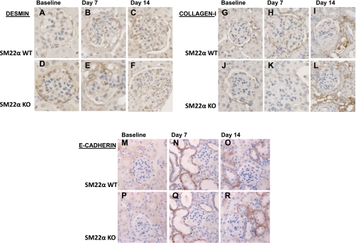
Fig. 12.
IHC for desmin, collagen type I, and E-cadherin in SM22α +/+ and SM22α −/− mice in experimental crescentic GN. Representative micrographs of IHC for intermediate filament protein desmin (A–F), interstitial matrix component collagen type I (G–L), and epithelial marker E-cadherin (M–R) are shown (original magnification ×40). At baseline, there is no significant difference in desmin staining between SM22α +/+ and SM22α −/− tissues (A and D). Following disease induction, there is increased desmin staining within the glomerular tuft area, with no significant difference between the two groups at the early (B and E) and late (C and F) time points. At baseline, there is no significant staining for collagen type I within the glomerular tuft area (G and J). Following disease induction, there is no significant increase in staining within the glomerular tuft area, but there is an increase in tubulointerstitial staining in both SM22α +/+ and SM22α −/− mice (H and K, I and L). At baseline, there is no significant staining in the glomerular tuft area for E-cadherin in SM22α +/+ and SM22α −/− mice (M and P). Following disease induction, there is increased tubular epithelial staining for E-cadherin, with no significant difference between the 2 groups at the early (N and Q) and late (O and R) time points.

