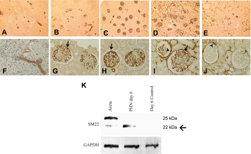
Fig. 2.
SM22α levels in passive Heymann nephritis (PHN) model of membranous nephropathy. A–J: representative micrographs of IHC for SM22α. Shown are control tissue (A and F) and tissue following disease induction at day 3 (B and G), day 6 (C and H), day 10 (D and I), and day 30 (E and J). In control tissue, only arterioles stained positive for SM22α. Following disease induction, there was a progressive increase in SM22α staining in glomeruli from days 3–10. By day 30, the immunostaining had decreased in the glomerular tuft, but remained prominent in parietal epithelial cells (PECs) and in the interstitium. Original magnification ×10 (A–E) and ×20 (F–J). Arrows indicate positive podocytes and arrowheads indicate positive PECs. K: Western blot for SM22α. In protein extracted from isolated normal glomeruli, no SM22α was detected. Protein extracted from isolated glomeruli of PHN rats showed abundantly expressed SM22α by day 6 of disease. Protein extracted from the aorta served as the positive control. GAPDH was used as a housekeeping protein to ensure equal protein loading. Arrow indicates band of interest at 22 kDa.

