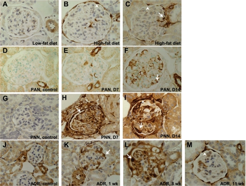
Fig. 3.
SM22α IHC in rodent disease models characterized by podocyte injury and proteinuria. Representative micrographs of IHC for SM22α. Original magnification ×40. Arrows indicate examples of positive podocytes; arrowheads indicate examples of positive PECs. A–C: diet-induced obesity (DIO) in mice. A: in mice fed a low-fat diet, there was no significant glomerular staining for SM22α. An adjacent vessel served as the internal positive control. B and C: in mice fed a high-fat diet, there was positive SM22α staining in both podocytes and PECs. D–F: puromycin aminonucleoside nephropathy (PAN) in rats. D: in rats that received vehicle alone, there was positive staining for SM22α in vessels only. E and F: by day 7 of disease, and increasing by day 14, there was de novo expression of SM22α in podocytes. G–I: passive nephrotoxic nephritis (PNN) in mice. G: in mice that received vehicle alone, SM22α staining was negative in glomeruli. H and I: by day 7, and continuing at day 14, there was de novo expression of SM22α, with positive staining in podocytes and PECs. J–M: adriamycin (ADR) nephropathy in mice. J: in mice receiving vehicle only, there was no significant staining in glomeruli. K: by 1 wk after administration of ADR, there was de novo expression of SM22α in podocytes and PECs. L: staining of SM22α increased by week 8 following ADR administration. M: by week 11 of disease, the SM22α staining of podocytes had diminished, but the staining in PECs remained strong.

