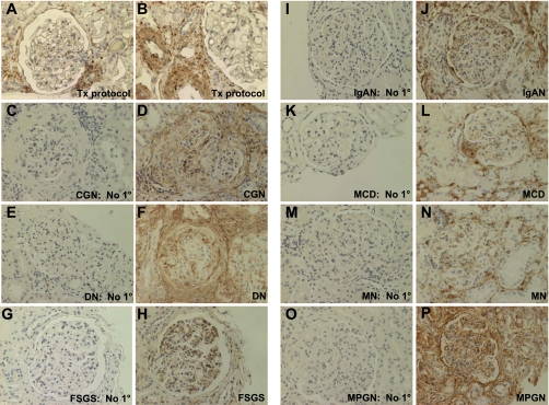
Fig. 4.
SM22α IHC in human diseases characterized by proteinuria. Representative micrographs of IHC for SM22α. A and B: protocol biopsy of transplant kidney tissue, considered “normal” control. There was no significant staining for SM22α within the glomeruli. The staining in vessels served as the internal positive control. C–P: in all cases, with omission of the primary antibody, there was no significant staining. In diseased tissues, there was positive intraglomerular staining for SM22α in a podocyte, PEC, and mesangial cell distribution. In some instances, there was marked periglomerular and tubulointerstitial staining associated with positively stained polymorphonuclear leukocytes within the interstitium. In addition, peritubular capillaries intensely stained positively for SM22α. C and D: collapsing glomerulopathy (CGN). E and F: diabetic nephropathy (DN). G and H: focal segmental glomerulosclerosis (FSGS). I and J: IgA nephropathy (IgAN). K and L: minimal change disease (MCD). M and N: membranous nephropathy (MN). O and P: membranoproliferative glomerulonephritis (MPGN).

