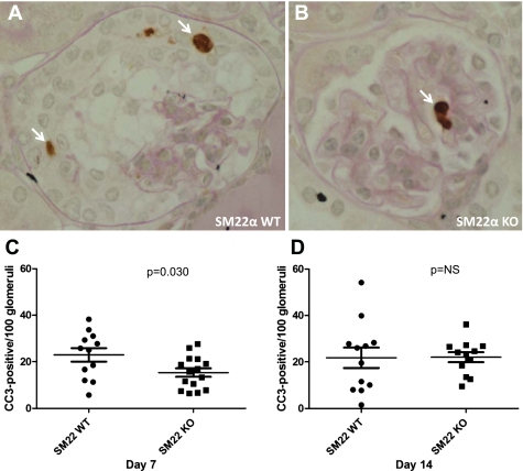
Fig. 7.
Apoptosis detection by IHC for cleaved caspase-3 (CC3) in SM22α +/+ and SM22α −/− mice following induction of crescentic GN. A and B: representative micrographs of CC3 staining in diseased glomerular tissue from SM22α +/+ mice (A) and SM22α −/− mice (B). Arrows indicate positively stained cells. C and D: quantification of positively stained cells for CC3 in tissues from diseased SM22α +/+ and SM22α −/− mice. C: at day 7 following disease induction, there were statistically significantly more cells staining positively for CC3 in SM22α +/+ tissue compared with SM22α −/− tissue (P < 0.05). D: by day 14, this difference was no longer statistically significant.

