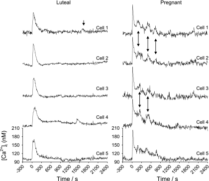
Fig. 3.
Synchronous [Ca2+]i bursts in neighboring cells in response to 100 μM ATP over 40 min. Multiple individual cell tracings from endothelium of a single intact UA from luteal NP (left) and pregnant (right) ewes are shown. After the initial peak, continued synchronous bursts by cells are as indicated by arrows.

