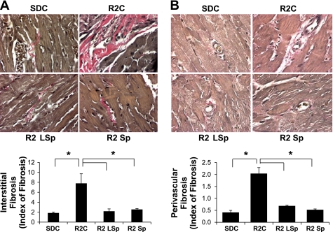
Fig. 6.
Markers of fibrosis for control and Sp-treated R2 rats. A and B: representative light micrographs (top) showing collagen fiber staining (pink) upon Verhoeff-van Gieson staining in the interstitium (A) and adventitia (B) surrounding coronary arterioles with bar graphs of relative degrees of fibrosis (bottom). *P < 0.05 vs. R2C rats.

