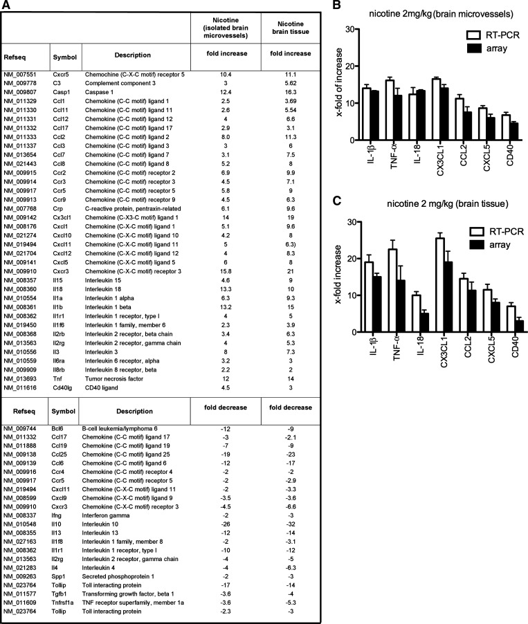
Fig. 1.
A: list of the transcripts modulated by nicotine in brain tissue and isolated brain microvessels from mice treated with nicotine (2 mg/kg) for 14 days. Fold increase/decrease indicate the level of up- or downregulation of transcripts compared with control (vehicle-treated mice). Three independent samples were analyzed by RT2 real-time PCR array. B and C: quantitative real-time PCR for IL-1β, TNF-α, IL-18, CX3CL1, CCL2, CXCL5, and CD40 was carried out on RNA from isolated brain microvessels (B) and brain tissue (C) from nicotine-treated (n = 3) and vehicle-treated mice (n = 3). Expression of target genes was normalized to control vehicle-treated mice. Values are presented as means ± SD. The fold changes obtained with RT-PCR were similar to those obtained by the PCR array.

