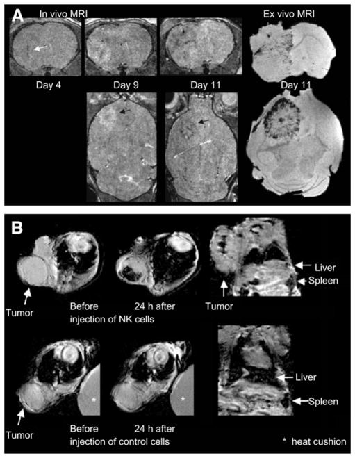
FIGURE 2.
(A) On left are 3-dimensional RARE images acquired after injection of labeled Sca1+ bone marrow cells showing hypointense regions within and around tumor, because of incorporation of labeled cells into vascular structures or within parenchyma of tumors; on right is ex vivo gradient-echo MR image at day 11. (Adapted with permission from (16). Research originally published in Blood [American Society of Hematology].) (B) At top are T2*-weighted images of HER2/neu+ NIH3T3 tumor (arrows) showing heterogeneous decline in signal intensity after injection of labeled NK cells directed against HER2/neu; at bottom, T2*-weighted images of HER2/neu+ NIH3T3 tumor (arrows) showing no change in signal intensity after injection of labeled nonspecific NK cells. (Adapted with kind permission of Springer Science and Business Media, from (18).)

