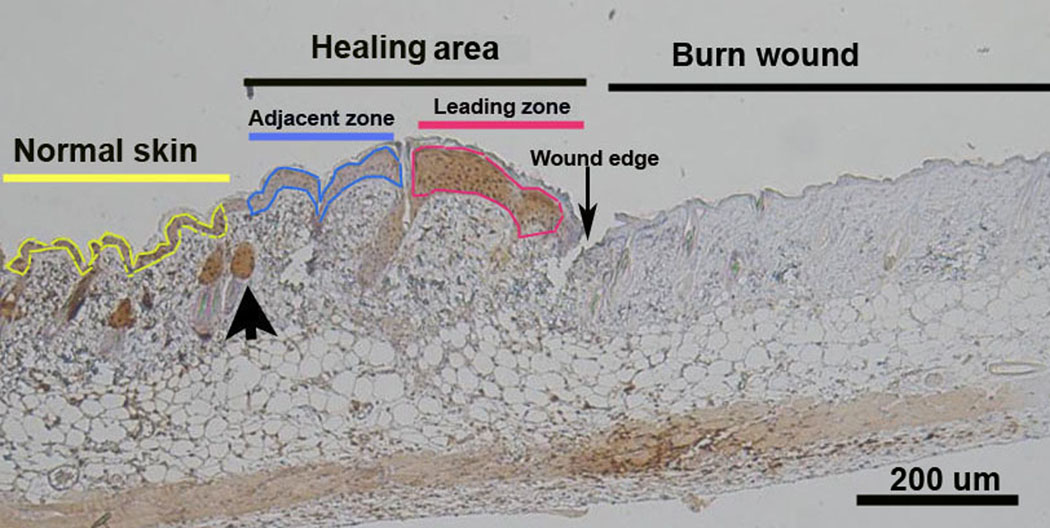
Figure 1.
Photomicrograph showing the location of hypoxia in the burn wound at day 4. To the right is coagulation necrosis of the burn wound. To the left is normal skin with hair follicles outlined in yellow. The area between the burn wound and the first normal hair follicle (large arrow) is defined as the Healing Area. The Healing Area has 2 portions: one is hypoxic and the other is not hypoxic. The Leading Zone outlined in red shows the pimonidazole positive brown staining indicating hypoxia. More proximal to the normal skin is the pimonidazole negative portion of the healing area termed the Adjacent Zone and outlined in blue.

