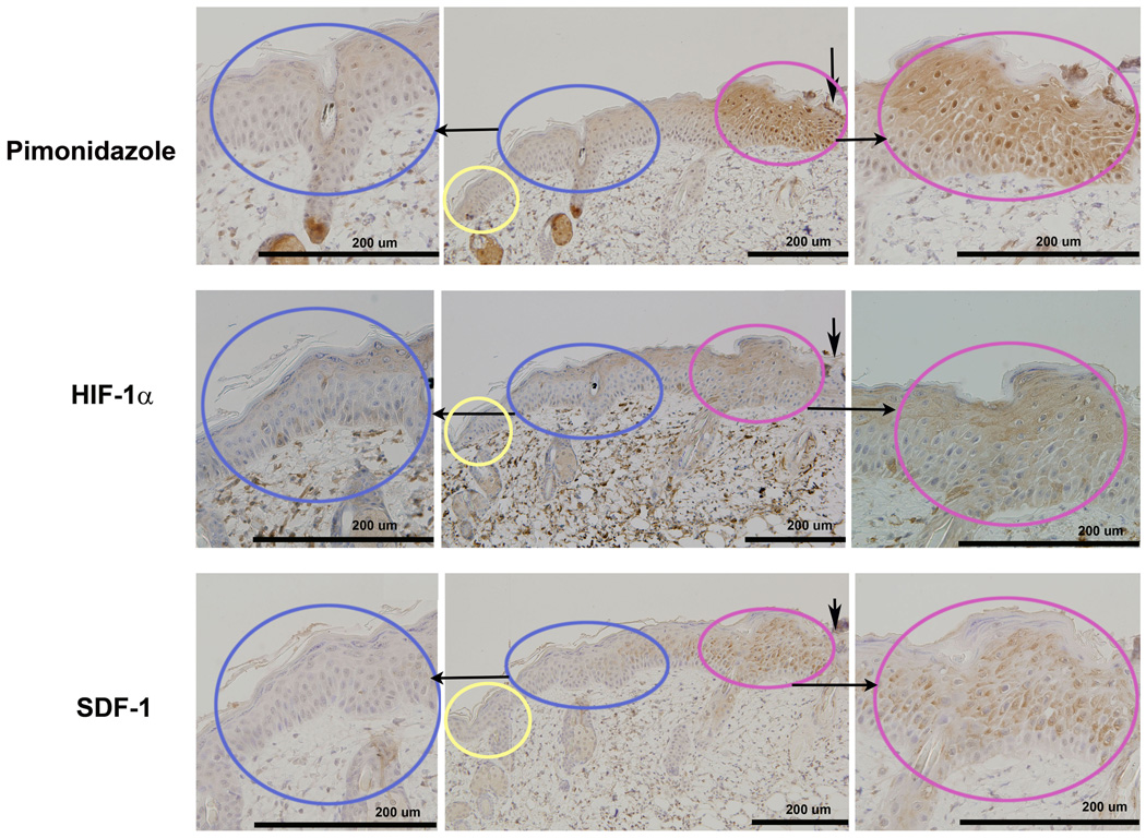
Figure 2.
Serial IHC sections show the localization of hypoxia, HIF-1 α, and SDF-1 in the leading zone: IHC of hypoxia (pimonidazole-1 staining, upper panel), HIF-1α (middle panel), and SDF-1 (lower panel) were carried out 72 hours after burn. The Leading Zone (in red) showed positive brown staining for all 3 markers. The Adjacent Zone (in blue) and normal skin (in yellow) did not stain positively for the markers. Serial sections from the same tissue block show the co-localized regions of the 3 stains.

