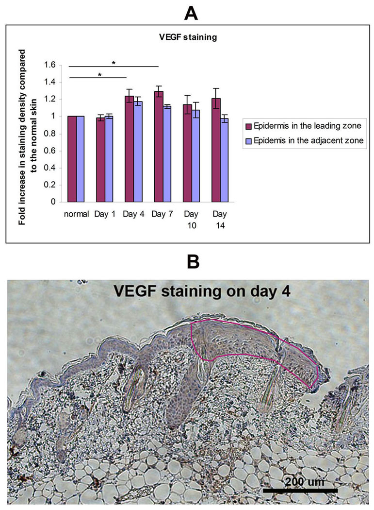
Figure 4.
VEGF in the Leading Zone: IHC analysis of VEGF in burn wound. (A) Staining density of the VEGF in Leading Zone, Adjacent Zone, and normal skin were quantitated using Image-Pro 5.1. Fold increase in staining intensity over normal skin was calculated (n=3 mice for each group, *P< 0.05, two-way ANOVA and Tukey post-tests. (B) Representative image of day 4 shown here. VEGF stains in brown.

