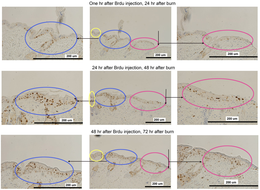
Figure 7.
In vivo BrdU pulse-and-trace experiment confirms the findings with Ki67. Mice were pulsed with BrdU at 48 hours after burn and traced at 1 hour (upper row), 24 hours (middle row) and 48 hours (lower row) after BrdU injection. At 1 hour after injection (2 days after burn), cells have a slightly increased staining in the adjacent zone as shown in the upper row. 24 hours later (middle row, 3 days after burn), rapidly proliferating cells are within the Adjacent Zone (in blue) but not in the Leading Zone (in red). 48 hours later (lower row, 4 days after burn), positively stained cells appear in the Leading Zone (in red) which indicates that the proliferating cells have migrated into the Leading Zone.

