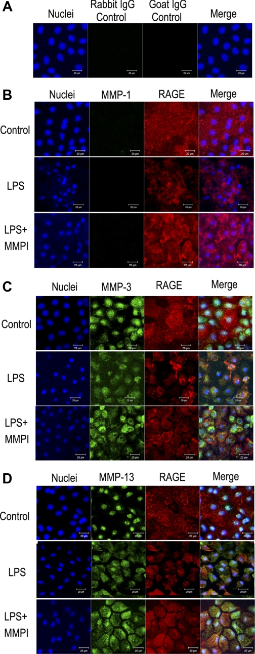
Fig. 4.
Expression of MMP-1 (B), MMP-3 (C), and MMP-13 (D) in the rat alveolar epithelial cells cultured on Transwell. Cells were also stained with anti-RAGE antibody (red), and nuclei are stained with DAPI (blue). A: results of negative control stain with rabbit IgG control and goat IgG control for anti-MMP antibodies (rabbit IgG) and anti-RAGE antibody (goat IgG). Each result was obtained as a representative result from consecutive 4 preparations in each study condition. And in each set of images, top row demonstrates control condition, middle row demonstrates cells treated with LPS (500 μg/ml), and bottom row demonstrates cells treated with LPS (500 μg/ml) and MMP-inhibitor 1 (MMPI, 200 μM) (LPS+MMPI), where MMPI was added to prevent loss of membrane-bound RAGE and detachment of cells. In control cells, MMP-3 and MMP-13 were expressed and their spatial expression overlapped with DAPI stain. In both LPS cells and LPS+MMPI cells, RAGE stain demonstrates loosening of cellular attachment and change in the spatial expression of MMP-3 and MMP-13. MMP-1 was negative in control cells, LPS cells, and LPS+MMPI cells.

