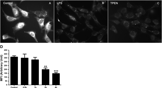
Fig. 1.
Effect of LPS on intracellular labile Zn in live sheep pulmonary artery endothelial cells (SPAEC) as determined by microspectrofluorimetry. SPAECs were control (A) or treated with LPS (100 ng/ml) (B) or N,N,N′,N′-tetrakis(2-pyridylmethyl)ethylenediamine (TPEN) (2 μM) (C) for 4 h. Cells were loaded with 5 μM FluoZin-3 AM and equal volume of Pluronic F-127 and imaged by epifluorescence microscope. The images represent fluorescence intensity of FluoZin-3-Zn complex in SPAECs. All images were captured with identical gain, 100% light intensity, 1-ms light exposure and 4 × 4 binning. D: time-dependent LPS-induced changes in FluoZin-3 fluorescence in live SPAEC. SPAECs were treated with HBSS (Ca2+/Mg2+) in the presence of LPS (100 ng/ml) for 30 min, 1 h, 2 h, and 4 h, respectively. Control cells received HBSS (Ca2+/Mg2+) in the absence of LPS for 4 h. The data represent means ± SE of mean fluorescence intensity (MFI) of 330–400 randomly selected cells from 5 experiments for each time point. Images were captured using identical gain and camera settings. For analysis of images, background illumination was subtracted from the readings. **P < 0.01 and ***P < 0.001, compared with control; 1-way ANOVA-Tukey.

