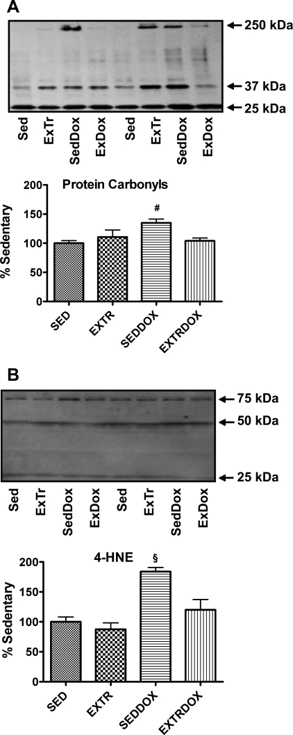
Fig. 2.
Protein carbonyls (A) and 4 hydroxy-2-nonenal (4-HNE; B) in soleus muscle were analyzed as indicators of oxidative damage. Representative Western blots are shown above graphs. Values (means ± SE) represent percent change. #Significantly higher than Sed and ExTrDox. §Significantly higher than Sed, ExTr, and ExTrDox.

