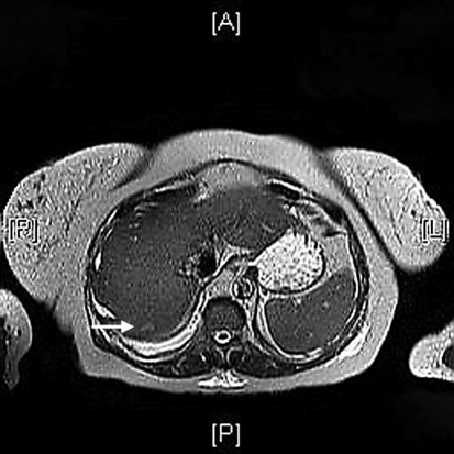
Fig. 1.
MRI abdomen with 11 mm nonspecific T2 hyperintense lesion in the posterior dome of the liver (arrow). Hepatosplenomegaly with smooth liver edges measuring 23 cm superior to inferior and the spleen 12.5 cm near the upper limit of normal. No changes suggestive of cirrhosis or fatty infiltration.

