Fig. 5.
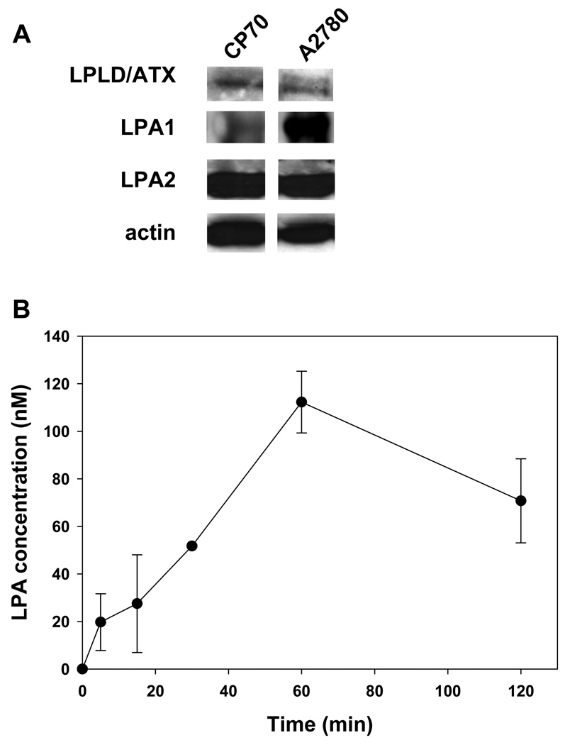
LPLD/ATX, LPA and LPA 1 and 2 are expressed in ovarian cancer cells A2780 and CP70. (A). A2780 and CP70 ovarian carcinoma cells were grown to sub-confluency. Shown is Western blot analysis of total protein from conditioning medium using anti-LPLD/ATX antibody and from cell lysates using antibody to LRA1, LPA2 and actin. (B). A2780 cells were irradiated with 3 Gy. LPA levels were measured in conditioning medium from A2780 cells at 0–120 minutes after irradiation using ELISA. Shown are average LPA concentration and SEM from three experiments.

