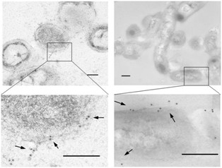
Figure 5.

Transmission electron micrographs of P. pneumotropica ATCC 35149 cells by immunoelectron microscopy with anti-rPnxIIIA IgG. Transmission electron micrographs of the P. pneumotropica ATCC 35149 cells that were first reacted with anti-rPnxIIIA IgG and then labeled with gold particles (10-nm) conjugated with rabbit IgG antibody. Arrows indicate the areas where gold labeling appeared on the cell surface. Left panel, cross-section of the bacterial cell. Right panel, longitudinal section of the bacterial cell. Bar = 0.2 μm.
