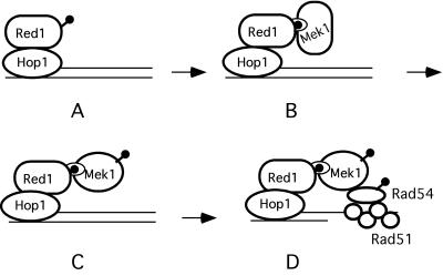
Figure 8.
Model for Hop1/Red1/Mek1 function during meiosis. (A) Hop1/Red1 complexes bind to DNA at sites where DSBs will form. (B) Phosphorylated Red1 forms a complex with the FHA domain of Mek1. (C) Mek1 is activated by phosphorylation of T327. (D) Mek1 phosphorylates substrates involved in the barrier to SCR. Lollipops indicate phosphates.

