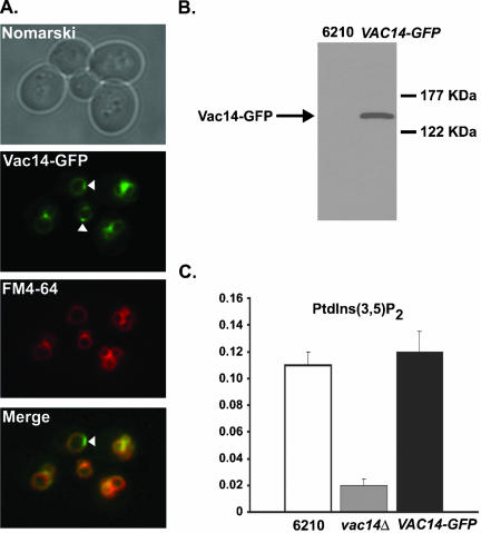
Figure 4.
Vac14 localizes to the limiting membrane of the vacuole. (A) Vac14-GFP–expressing wild-type cells were labeled with the fluorescent dye FM4-64 to visualize the vacuole membranes. The localization of FM4-64 and Vac14-GFP were compared by fluorescent microscopy. (B) The total cellular protein content of wild-type and wild-type expressing Vac14-GFP were TCA-precipitated and resolved by SDS-PAGE. Vac14-GFP was detected by Western blotting using anti-GFP antibody. (C) Quantitation of PtdIns(3,5)P2 in wild-type, vac14Δ, and VAC14-GFP strains. 3H-labeled phosphoinositides were isolated, resolved and measured as described in MATERIALS AND METHODS. The levels of PtdIns(3,5)P2 are expressed as a percentage of the total 3H-labeled phosphoinositides analyzed by HPLC.

