Figure 5.
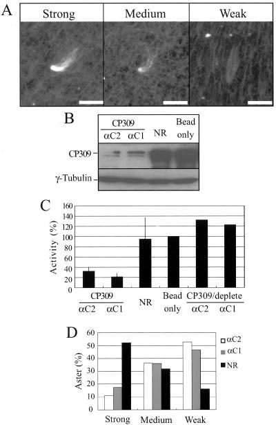
CP309 is required for microtubule nucleation from centrosomes in vitro. (A) Three typical asters with strong, medium, and weak microtubule nucleation are shown. Bars, 5 μm. (B) Depletion of CP309 from embryo extracts. Embryo extracts were depleted using two affinity-purified antibodies (αC1 or αC2) against different regions of CP309, mock-depleted using nonimmunized rabbit IgG (NR), or with beads alone. Western blotting analysis showed that 70–90% of the CP309 was depleted from embryo extracts, whereas the amount of γ-tubulin was unchanged. (C) Centrosome-complementing assays. Extracts were immunodepleted with NR, beads alone, or αC1, αC2, or αC1 and αC2 that were depleted of CP309-specific antibody. Each of these extracts was then used to reconstitute centrosomes from KI extracted centrosome scaffolds. Asterforming activity was quantified as described above and expressed as percentage of the mock-depleted embryo extracts (beads only). Error bars represent SD from four independent experiments. (D) Percentages of asters with strong, medium, or weak microtubule nucleation in the extracts depleted with NR, αC1, or αC2.

