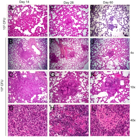
Figure 4. Histopathology of lungs of rats infected with M. tuberculosis W4, H&E.
A, E and I, inoculum 103 CFU, magnification 10×; A. Day 14 post infection. Alveolar cell wall thickening is observed. E. Day 28 post infection. Small cellular aggregates are seen. I. Day 60 post infection. Small granulomas have formed. B, C and D - inoculum 104 CFU, 14 days post infection. B. Small peribronchial cellular aggregates are observed, magnification 4×. C. Magnification 10× and D, The lesions consist of mixed lymphocytes, PMNs and macrophages, magnification 40×. F, G and H - inoculum 104 CFU, 28 days post infection; magnification 4×, 10× and 40× respectively. Small, well organized granulomas with central area of macrophages and cuff of lymphocytes. J, K and L - inoculum 104 CFU, 60 days post infection; magnification 4×, 10× and 40× respectively. Well structured granulomas with differentiated macrophages.

