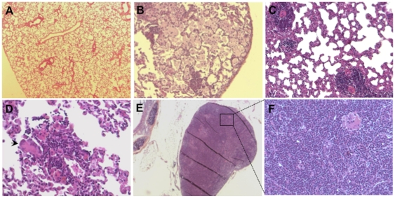
Figure 5. Histology of lungs from rats infected with W4 for 180 days.
(A) Normal appearing lung parenchyma, magnification 6.5×. (B) Lung with variable numbers of foamy macrophages, magnification 10×. (C) Small granulomas containing lymphocytes and epithelioid cells, magnification 20×. (D) A small inflammatory cell aggregate containing a large numbers of lymphocytes and a multinucleated giant cell, magnification 40×. (E) An infiltrated mediastinal lymph node with lymphocytes and epithelioid histiocytes, magnification 6.5×. (F) Same as E, magnification 20×.

