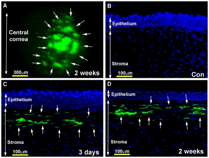
Figure 1. Representative in vivo fluorescence stereomicrograph (A) and tissue sections (B–D) of rabbit corneas showing AAV5-mediated GFP gene expression at 3-day and 2-week time points.
Topical application of AAV5-GFP vector selectively transduced anterior keratocytes (arrows) located beneath the epithelium (C, D). No transgene expression was detected in control corneas (A). The rabbit corneas collected at 4-week and 16-week showed similar levels of GFP expression with immunostatining (data not shown). Nuclei are stained blue with DAPI. Scale bar denotes 100 µm.

