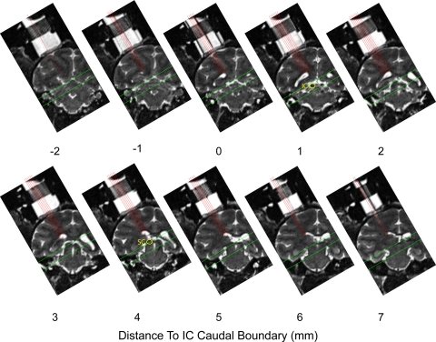
Fig. 1.
Locations mapped in monkey A illustrated on MR images. A series of coronal MR images spanning the 10- mm range that was sampled physiologically. Voxels were 0.5-mm cubes. Images were rotated into the plane of recording by placing electrodes in the recording grid, visible ∼0 and 7 mm. Each panel corresponds to a single mediolateral row of grid locations at a given position in the anterior/posterior dimension (interleaved coronal slices are not displayed). Red lines indicate the approach of each of the recording penetrations in the medial/lateral dimension. Green lines indicate the targeted area; recordings shallower and deeper than these borders were discarded. Locations of the inferior colliculus (IC) and superior colliculus (SC) are indicated on 2 of the panels.

