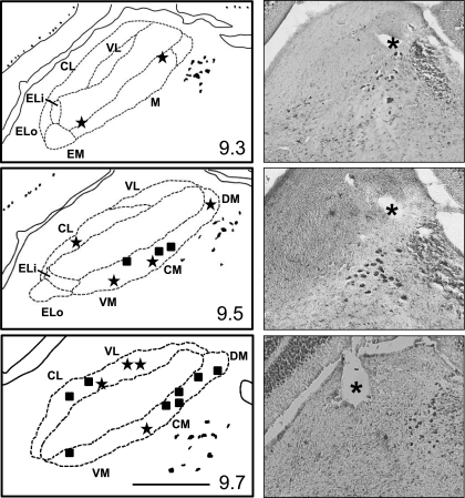
Fig. 5.
Histological results showing recording site for 21 cells. Left: line drawings of coronal sections at various AP levels through the PbN. Numbers in bottom right of each drawing indicate distance in millimeters caudal to bregma. Line in bottom right of bottom drawing indicates 0.5 mm. DM, dorsomedial n.; CM, central medial n.; VM, ventromedial n.; VL, ventral lateral n.; CL, central lateral n.; ELo, external lateral outer n.; external lateral inner n. Right: photomicrographs of coronal sections showing lesions (asterisks) marking recording sites at AP levels corresponding to the line drawings to the left.

