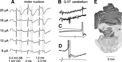
Fig. 3.
Comparison of IPSPs evoked in a dh DSCT neuron by stimuli of different intensities. A: IPSPs evoked in the dh DSCT neuron illustrated in Fig. 2B by changing intensity of stimuli applied in the posterior biceps-semitendinosus (PBST) motor nucleus at the depth of 2.4 mm. Averages of 10 records are shown. Dotted lines indicate onset of IPSPs evoked by the third 20 and 10 μA stimuli. The figures below the records indicate their latencies; note difference of 0.8 ms. B–D: records identifying this neuron as the spinocerebellar neuron. B: records obtained just before the penetration illustrating collision between spike potentials evoked by stimulation of the cerebellum and of the quadriceps (Q) nerve; spikes from the cerebellum are seen to the right (top traces) only when they were not preceded by the synaptically evoked spikes (those in the middle of the lower traces). C and D: intracellularly evoked antidromic spike potentials following stimulation of the cerebellum and synaptically evoked spikes induced in the same neuron by stimulation of Q. E: reconstruction of 3 stimulation sites in the cerebellum in approximately the same plane; the arrow indicates the right nucleus interpositus.

