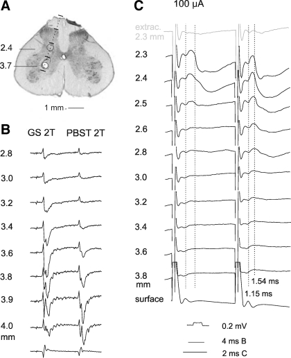
Fig. 8.
An example of postsynaptic potentials evoked from outside but not from within motor nuclei in SCT neurons. A: an electrode track along which stimuli were applied while recording from SCT neurons. Upper circle shows the location of an electrolytic lesion made at the depth at which maximal antidromic field potentials were evoked from the gastrocnemius-soleus nerve. Lower circle shows the area from which PSPs were most efficiently evoked. B: records of field potentials evoked at different depths (indicated to the left) by stimulation of gastrocnemius-soleus (GS) and posterior biceps-semitendinosus (PBST) nerves. C: intracellular records from a SCT neuron in which EPSPs and IPSPs were evoked by 100 μA from the depths 2.3–2.8 mm but not from within motor nuclei. Note that the most effective stimuli were those applied at the 2.3–2.4 mm depths, as judged both by the slopes of the rising face of the IPSPs and effects of not only the second but also the first stimuli. Bottom records in B and C are from the surface of the spinal cord. Top record in C is from just outside the neuron. Dotted lines in C indicate onset of EPSPs (latency 1.1–1.2 ms) and IPSPs (latency 1.5–1.6 ms).

