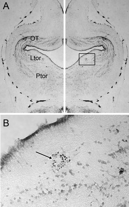
Fig. 2.
A: cresyl-violet-stained horizontal section through a brain that was lesioned by direct current at the end of the final ventral-going electrode track, marking the recording site of a cell nonselective for click rate. The left half of the figure is a reverse of the right, for the purposes of nuclei identification. Penetration was perpendicular to the plane of section. Ltor, laminar nucleus of the torus semicircularis (TS); OT, optic tectum; Ptor, principal nucleus of the torus semicircularis. B: enlargement of the box in A showing red blood cells that mark the lesion location in the laminar nucleus of the TS.

