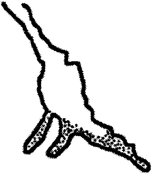
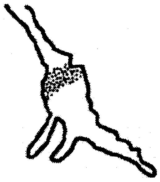
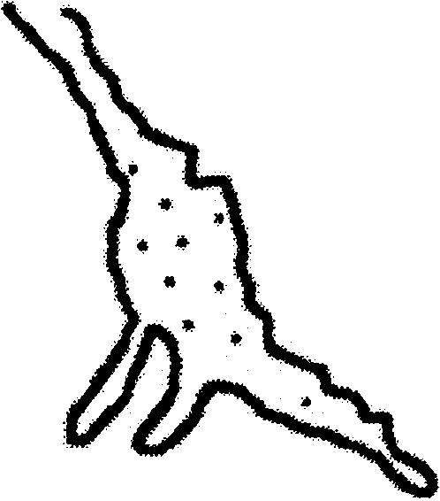
Table 2.
Quantification of Mena distribution in the axonal growth cone
| Mena localization at the growth cone | Control | ARNO-wt | ARNO-E156K | Rac1-N17 |
|---|---|---|---|---|
| At the leading edge lamellae and filopodia | 100 (16) | 100 (16) | 0 | 100 (8) |
 |
||||
| At the base of the growth cone | 0 | 0 | 31 (5) | 0 |
 |
||||
| Depleted from the growth cone | 0
|
0
|
69 (11)
|
0
|
 |
Hippocampal neurons were transfected with myc-tagged ARNO wild type, ARNO-E156K, and FLAG-tagged Rac1-N17, labeled with anti-Mena antibody and visualized by deconvolution microscopy. Quantification is expressed as percentage of transfected cells (number of cells). Mena was localized at the leading edge lamellae and filopodia in untransfected cells as well as in cells overexpressing either wild-type ARNO or Rac1-N17, at the base or proximal region of the growth cone in cells expressing ARNO-E156K, or depleted from the growth cone in cells expressing ARNO-E156K. n = 16 or 8.
