
Figure 5. Active site stereo views of crystallographically characterized apo-CFPs illustrating possible heme environments.
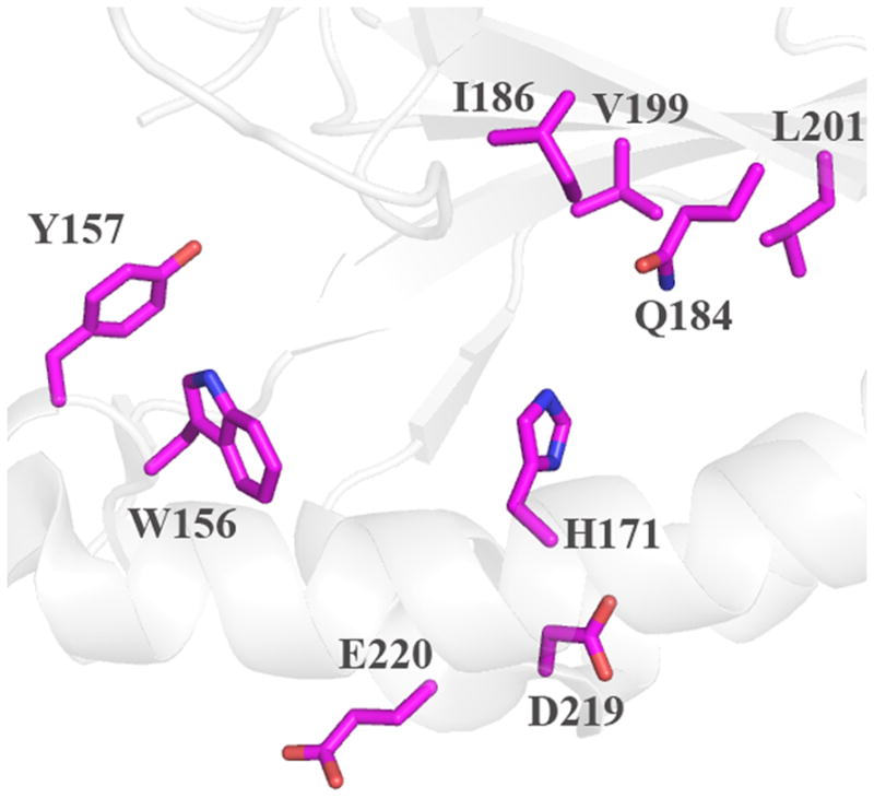
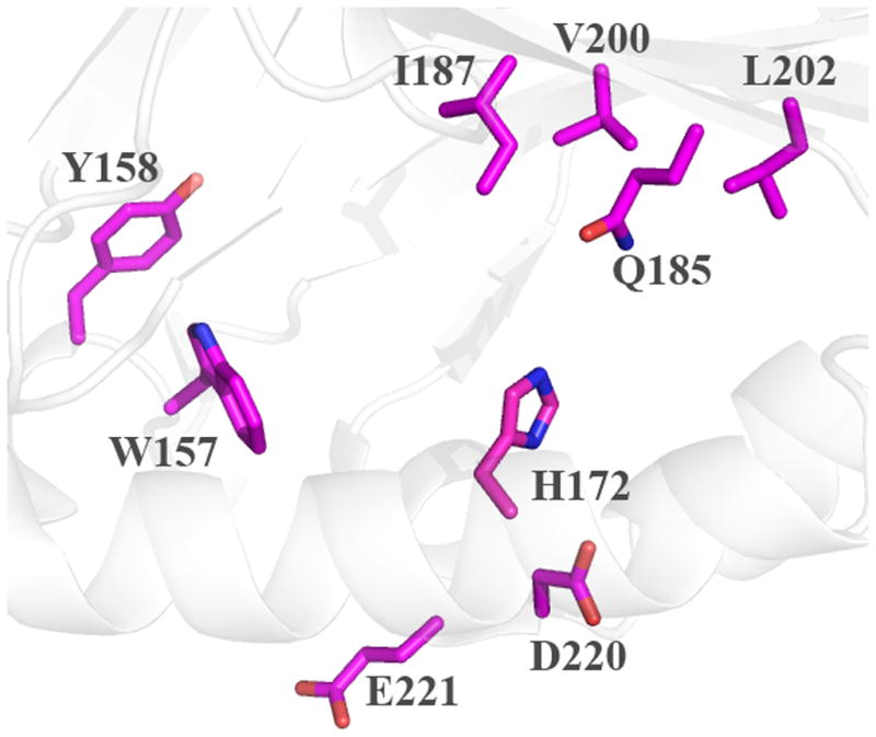
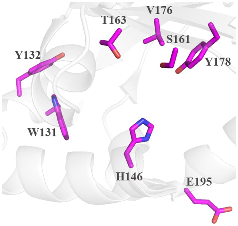
Cartoon diagram representations of protein monomers (carbon light grey) are shown, with side-chains of active site residues shown as sticks. Residues lining the expected heme pocket that fall within strictly conserved secondary structure elements are colored by atom (carbon magenta). Note that the same residues were also identified from primary sequence alignments with DA-Cld (Table 1). (A) G. stearothermophilus (Firmicutes); (B) T. thermophilus (Deinococcus-Thermus). (C) T. acidophilum (Euryarchaeota). This figure was generated using PyMOL (http://www.pymol.org/).