
Figure 6.
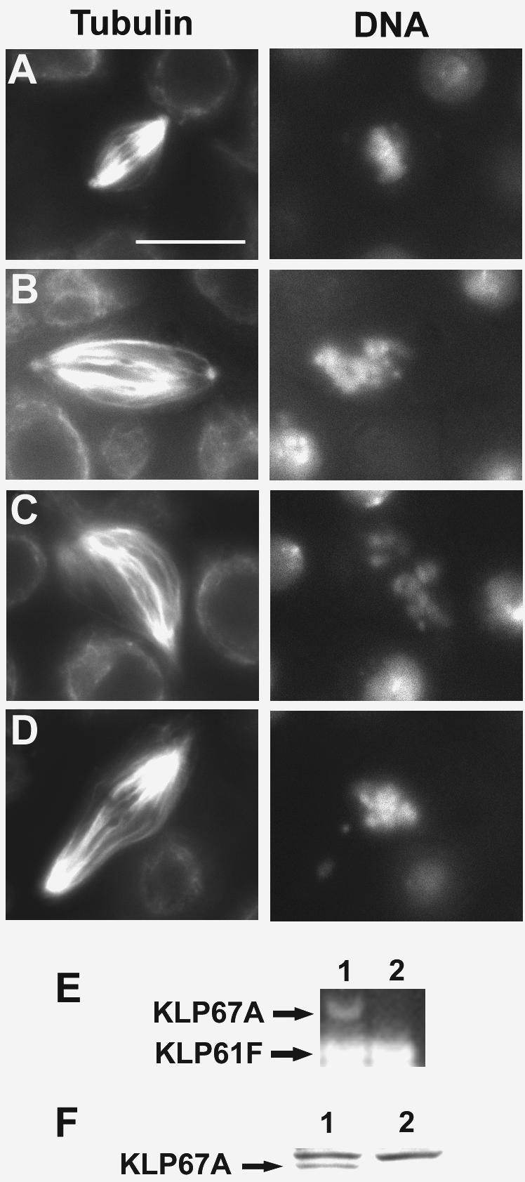
Klp67A dsRNA treatment in DL2 cells results in elongated spindles and mitotic arrest. (A–D) Cytological phenotypes caused by KLP67A depletion. Klp67A (RNAi) cells and control cells were fixed and stained for α-tubulin and DNA (with DAPI). (A) Mock-transfected control cells. (B–D) Cells treated with Klp67A dsRNA. (A) Metaphase. (B–D) Metaphase-like spindles in RNAi cells. Bar, 10 μm. (E) Semiquantitative RT-PCR assay shows undetectable levels of Klp67A mRNA in dsRNA-treated samples. 1, control; 2, cells treated with Klp67A dsRNA. Klp61F mRNA amplified by RT-PCR was used as an internal control. (F) Western blot analysis of DL2 cells treated with Klp67A dsRNA. Forty micrograms of protein was loaded in each lane, and the blot was probed with a rabbit polyclonal antibody to KLP67A. The common background band serves as a loading control.