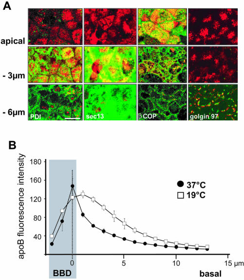
Figure 2.
Characterization of apoB-containing subcellular compartments in Caco-2 cells. (A) Immunolocalization of apoB (red channel) at the apex, and 3 μm and 6 μm below the apical plane in Caco-2 cells, compared with that for PDI (endoplasmic reticulum), sec13 (COPII vesicles), β-Cop (COPI vesicles), and Golgin 97 (trans-Golgi) (green channels). Bar, 10 μm. (B) Effect of temperature on the distribution of apoB; cells were cultured at 37°C or 19°C for 3 h. The localization of apoB was determined by confocal microscopy with respect to the apical domain (BBD, gray area), which was labeled with nonpermeant reactive biotin (sulfo-NHS-Biotin) detected with tetramethylrhodamine B isothiocyanate-conjugated streptavidin. For each area that was analyzed, point zero corresponds to the maximal intensity of the biotin labeling of the brush-border membrane (dashed line). The graphs represent the intensity of apoB-associated fluorescence that was evaluated for 12 areas composed of nine to 11 cells at 1-μm intervals along the Z-axis in the BBD and from point 0 to the basal part of the cells.

