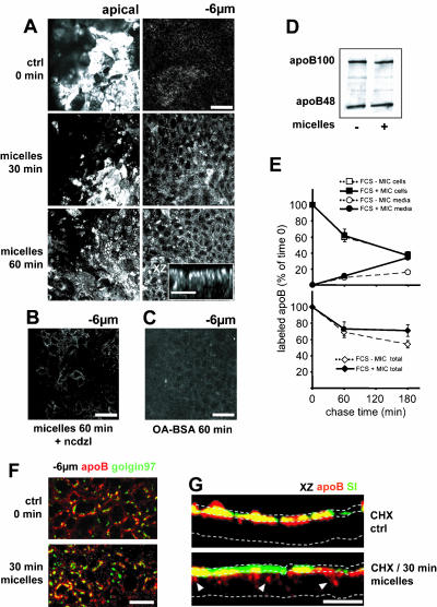
Figure 6.
apoB traffic after the apical application of micelles. (A) apoB immunolocalization at the apex and 6 μm below the apex, 30 and 60 min after the addition of lipid micelles; the inset in the bottom panel represents an XZ projection of the XY acquisitions 60 min after micelle delivery. (B) apoB immunolocalization 6 μm below the apex 60 min after micelles delivery in the presence of nocodazole (ncdzl). (C) apoB immunolocalization 6 μm below the apex 60 min after the addition of BSA-complexed oleic acid (OA-BSA). Bars, 50 μm; for inset in A, 10 μm. (D) Western blot analysis of apoB distribution in Caco-2 cells cultured with FCS-containing medium in the basal compartment in the presence (+) or the absence (–) of apical micelles supply. Forty micrograms of proteins were loaded in each lane. (E) Pulse-chase experiments of apoB in Caco-2 cells cultured with FCS-containing medium in the basal compartment in the presence or absence of apical micelles supply. After 1 h of labeling with [35S]Met/Cys (0) and at 1 and 3 h of chase, apoB was immunoprecipitated from cell extracts (200 μg of proteins for each condition) and media (250 μl in each condition), electrophoresed, analyzed by fluorography, and counted. Results, from four independent determinations, are expressed as the percentage of labeled apoB recovered in cells and media compared with the 100% amount of labeled apoB at t = 0 of chase, i.e., 15,700 ± 850 and 16,500 ± 1,300 cpm/mg proteins in the presence or absence of apical micelles supply respectively. (F) Immunolocalization of apoB (red channel) and Golgin 97 (green channel) 6 μm below the apical plane in Caco-2 cells without (top) or with lipids micelles in the medium for 30 min (bottom). Bar, 10 μm. (G) XZ representations of apoB (red channel) and SI (green channel) immunolocalization in Caco-2 cells treated with CHX without (top) or with (bottom) incubation with lipids micelles for 30 min. Dotted lines delimit the brush-border domain and the basal level of Caco-2 cells in the XZ representation. White arrowheads indicate delocalized apoB. Bar, 20 μm. Note that the apical application of micelles specifically induced rapid vesicular traffic of apoB from apical to lateral perinuclear areas.

