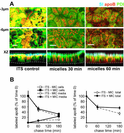
Figure 7.
The delivery of micelles to Caco-2 cells cultured in ITS medium induces the export of apoB from the ER and the completion of its traffic. (A) Immunolocalization of apoB (red channel), SI (blue channel) and PDI (green channel), at 3 μm and 6 μm below the apical plane and in an XZ representation of Caco-2 cells cultured in ITS medium before (ITS control) and after 30 and 60 min of incubation with micelles. Dotted lines delimit the brush-border domain and the basal level of Caco-2 cells in the XZ representation. Bars, 10 μm. (B) Pulse-chase experiments of apoB in Caco-2 cells cultured with ITS-containing medium in the basal compartment in the presence or absence of apical micelles supply. After 1 h labeling with [35S]Met/Cys (0) and at 1 and 3 h of chase, apoB was immunoprecipitated from cell extracts (200 μg of proteins for each condition) and media (250 μl in each condition), electrophoresed, analyzed by fluorography, and counted. Results, from four independent determinations, are expressed as the percentage of labeled apoB recovered in cells and media compared with the 100% amount of labeled apoB at t = 0 of chase, i.e., 16,500 ± 900 and 18,000 ± 1,700 cpm/mg proteins in the presence or the absence of apical micelles supply, respectively.

