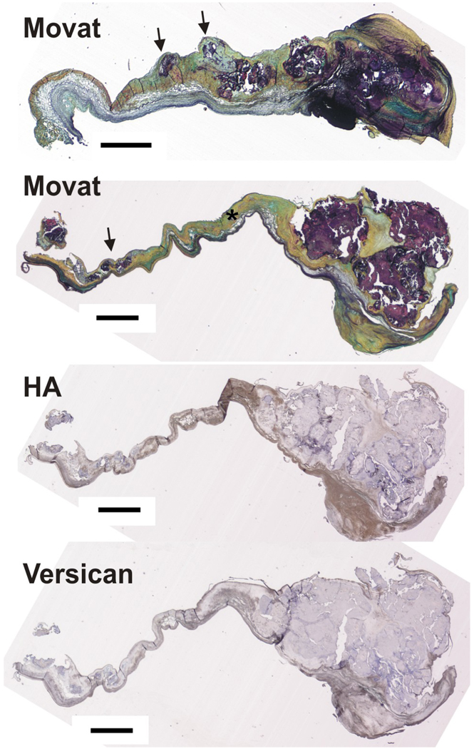
Figure 1.
Upper two images: Movat pentachome stain of two calcified aortic valves showing large nodules at the distal end and small prenodules (indicated by arrows) more proximal to the annular edge of the leaflet. Asterisk indicates normal fibrosa. Lower two images: one of the same calcified valves stained for hyaluronan (HA) and versican. Scale bar = 1 mm.

